Fig. 7.
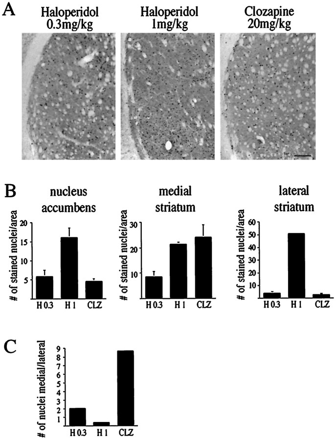
All treatments (haloperidol 0.3 and 1 mg/kg, clozapine 20 mg/kg) induce Fos levels in the striatum with variations in anatomical distribution. A, Fos protein staining in the lateral striatum after treatment with haloperidol (0.3 and 1 mg/kg) or with clozapine (20 mg/kg). No Fos-positive nuclei were observed after treatment with saline (data not shown). Scale bar, 0.25 mm.B, Comparison of the number of Fos-positive nuclei in the nucleus accumbens, the medial striatum, and the lateral striatum after treatment with haloperidol (0.3 and 1 mg/kg, H 0.3, H 1) and with clozapine (20 mg/kg, CLZ).n = 3 for H 1 and CLZ, n = 5 for H 0.3 (see also Fig. 8). Bars in graphs present the average fold induction ± SEM of six striatal areas of three rats treated with haloperidol (1 mg/kg) or clozapine (20 mg/kg), or five rats treated with haloperidol (0.3 mg/kg). C, The ratio of Fos-positive nuclei in the medial over the lateral striatum after treatment with haloperidol (0.3 and 1 mg/kg) and clozapine (20 mg/kg). The medial striatum was particularly sensitive to clozapine and 0.3 mg/kg haloperidol, whereas the lateral striatum responded strongly to 1 mg/kg haloperidol.

