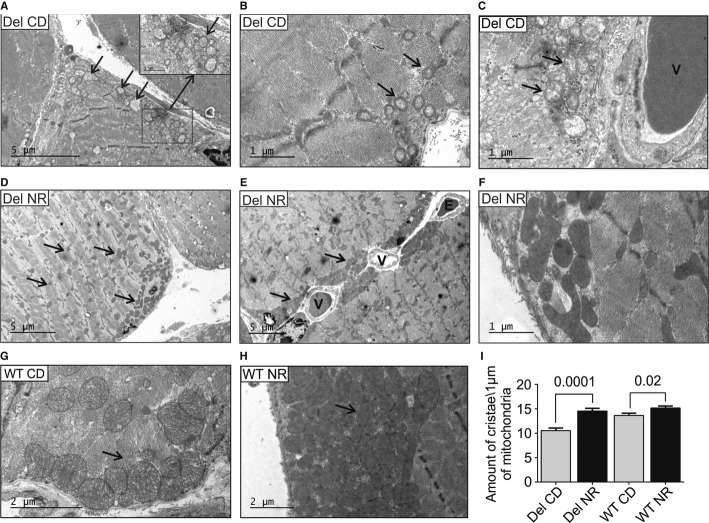
Figure 2. NR induces skeletal muscle mitochondrial mass and prevents the development of mitochondrial ultrastructural abnormalities in mitochondrial myopathy.
- A–C Deletor quadriceps femoris muscle ultrastructure on chow diet shows increased amount of swollen mitochondria with concentric crista abnormalities (arrows) especially in subsarcolemmal regions.
- D–F Deletors on NR diet. Mitochondria with normal ultrastructure and dense matrices are prominent subsarcolemmally (D–F), especially around the vessels (E, arrows), and also intramyofibrillar mitochondria show increased volume and matrix density (D, arrows). NR-fed Deletors show high crista density (F).
- G WT mice on CD, showing subsarcolemmal mitochondria (arrow).
- H WT mouse on NR diet with remarkable accumulation of subsarcolemmal mitochondria with dense matrices, and high crista density.
- I Quantification of mitochondrial crista density in Deletors and WT mice on CD or NR diets. In image analysis, a “measuring stick” of 1 μm, placed perpendicular to the crista lamellae, was used to count crista content. A total of 20 μm of mitochondrial length was calculated per each sample electron micrograph. n = 3 in each group.
Data information: Numbers above columns indicate P-values. All values are presented as mean ± s.e.m. (Student's t-test). Bars in electron micrographs indicate magnification. Del, Deletor; WT, wild-type mice; CD, chow diet; NR, nicotinamide riboside; V, blood vessel; E, erythrocyte.

