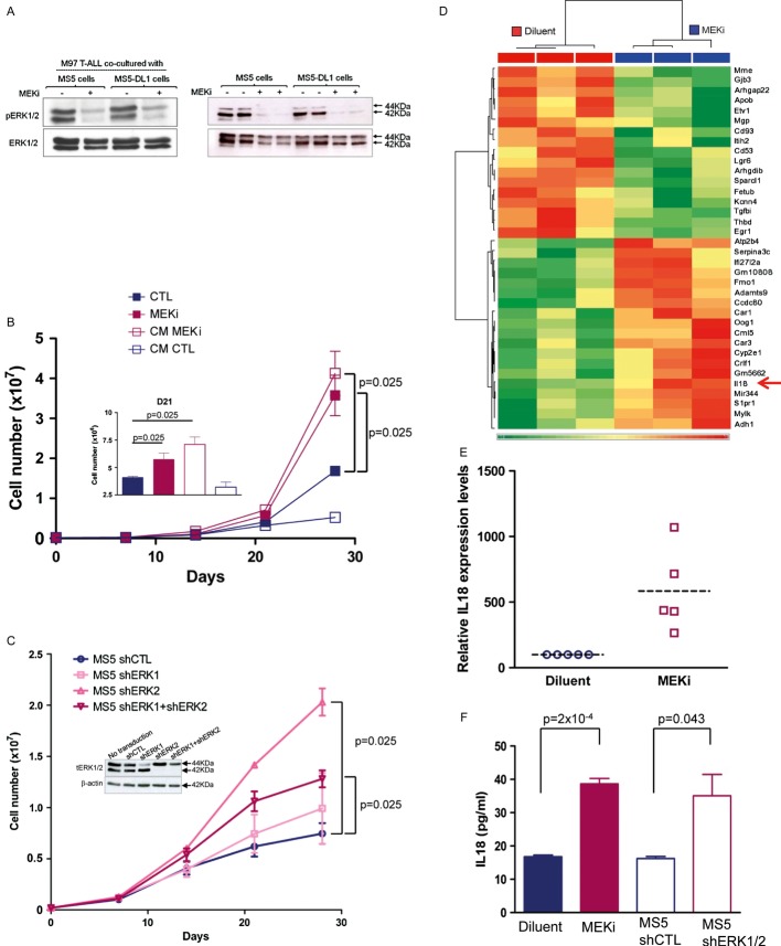
Figure 2. MEKi induces IL-18 production by MS5 stromal cells.
- Western blot analysis of protein lysates from M97 T-cell acute lymphoblastic leukaemia (T-ALL) cells co-cultured with MS5 or MS5-DL1 (left panel) and from MS5 or MS5-DL1 cells (right panel) incubated (+) or not (−) with the MEKi PD184352 for 1 h before harvesting.
- Assessment of M18 T-ALL cell proliferation when cultured in the presence of conditioned medium (CM) from control (DMSO) (CM CTL) or MEKi-treated (1 μM PD184352, CM MEKi) MS5 cells. Data are representative of three experiments performed in triplicate. Inset shows the details of day 21 (mean ± s.d.; Mann–Whitney non-parametric test).
- Proliferation of M69 T-ALL cells co-cultured with shERK1- and/or shERK2-transduced MS5 cells (inset shows the reduction of ERK1/2 protein expression following transduction with the different short hairpin RNAs). Data are representative of three experiments performed in triplicate (mean ± s.d.; Mann–Whitney non-parametric test). tERK1/2, total ERK1/2 proteins.
- Microarray analysis (Affymetrix arrays) of MS5 cells cultured in the presence of 1 μM PD184352 (MEKi) or diluent for 7 days. Heatmap shows the transcripts, the expression of which was most significantly different in the two conditions (arrow indicates IL-18).
- IL-18 mRNA expression in MS5 cells grown in the presence of 1 μM PD184352 or not (diluent) for 1 week (mean ± s.d. of five independent experiments).
- IL-18 protein expression in the supernatants of MS5 cells treated with 1 μM PD184352 or diluent for 1 week and in the supernatants of MS5 cells 1 week after transduction with shERK1+2 or shCTL (mean ± s.d. of triplicates; Mann–Whitney non-parametric test).
Source data are available for this figure.

