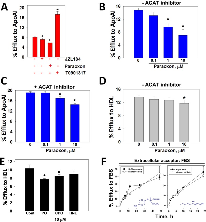
Figure 3.
Effect of xenobiotic and lipid electrophile HNE treatments on macrophage cholesterol efflux. (A) Macrophages loaded with acLDL/[3H]-cholesterol were treated with vehicle (ethanol), JZL184 (1 μM), paraoxon (1 μM), or synthetic LXR ligand T0901317 (10 μM) in the presence of ApoA1, and the extent of [3H]-cholesterol efflux after 24 h was determined. [3H]-Cholesterol efflux from macrophage foam cells to ApoA1 was determined as a function of paraoxon concentration in the absence (B) or presence (C) of ACAT inhibitor. (D) [3H]-Cholesterol efflux from macrophage foam cells to HDL was determined as a function of paraoxon concentration in the absence of ACAT inhibitor. (E) The extent of [3H]-cholesterol efflux from macrophages loaded with acLDL/[3H]-cholesterol and treated with either ethanol (control), paraoxon (PO), chlorpyrifos oxon (CPO), or 4-hydroxynonenal (HNE) in the presence of HDL was determined. (F) Time-course of [3H]-cholesterol efflux from acLDL/[3H]-cholesterol loaded macrophages treated with toxicants. Cells were treated with either PO or HNE (10 μM) for 24 h without cholesterol acceptors, followed by a 0–48 h efflux period with 10% v/v fetal bovine serum serving as the cholesterol acceptor. The toxicants were present in the culture media throughout the efflux period. Chemical structures for PO and HNE are indicated in graphs. Data in each panel represent the mean ± SD of 3 dishes; * p < 0.05, one-way ANOVA followed by Dunnett’s test.

