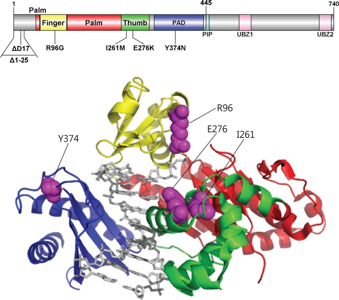
Figure 1.
Locations of genetic pol ι variations. Structure of human pol ι(26–445) (PDB code, 2FLL) bound to primer/template DNA and incoming nucleotide is shown using Pymol. Pol ι(26–445) is shown in cartoon ribbons, and the primer/template DNA and nucleotide are shown in gray sticks. The finger, palm, thumb, and PAD domains are colored yellow, red, green, and blue, respectively. The amino acid residues (in purple spheres) of genetic pol ι variants are indicated. The structural domains of pol ι are shown in the upper schematic diagram using DOG (version 2.0),60 where positions of amino acids related to six studied variations are indicated.

