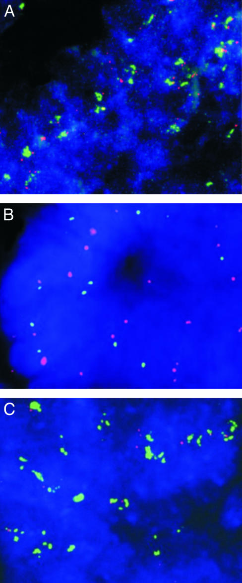
Fig. 3.
TYMS amplification assessed by interphase FISH. Analysis of interphase nuclei from a colorectal cancer metastasis to the liver after 5-FU treatment (A). Matched colorectal adenoma obtained before 5-FU treatment (B) and colorectal cancer obtained after 5-FU neoadjuvant treatment (C) from a patient with familial adenomatous polyposis. Nuclei are visualized with 4′,6′-diamidino-2-phenylindole stain (blue); TYMS probe (located on chromosome 18p, 0.8-1.0 Mb from the telomere) is visualized by using FITC-avidin (green), and chromosome 18 control probe (located on chromosome 18p, 13.0-13.2 Mb from the telomere) is visualized by using tetramethylrhodamine B isothiocyanate-conjugated antibodies (red). Increased TYMS gene copy number was observed only in patients previously treated with 5-FU (A and C). (Magnification: ×600.)

