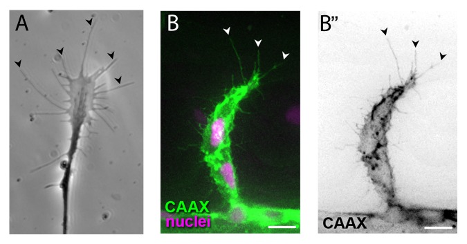
Figure 2. Axonal growth cones (A, image courtesy of Isabelle Brunet) and endothelial tip cells (B) extend long filopodia (arrowheads) in the direction of migration. Scale bars, 10µm.

Figure 2. Axonal growth cones (A, image courtesy of Isabelle Brunet) and endothelial tip cells (B) extend long filopodia (arrowheads) in the direction of migration. Scale bars, 10µm.