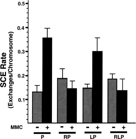
Figure 4.
Sister chromatid exchange assay in mutant MEFs. SCE rate determined in BrdU-labeled, acridine-orange-stained MEF cell metaphase spreads of the indicated genotypes. (P) p53-/-; (RP) Rad54-/- p53-/-; (LP) Lig4-/- p53-/-; (RLP) Rad54-/- Lig4-/- p53-/-; (P + MMC) p53-/- cultured in the presence of MMC, as a positive control for induced SCE. Error bars represent S.E.M.

