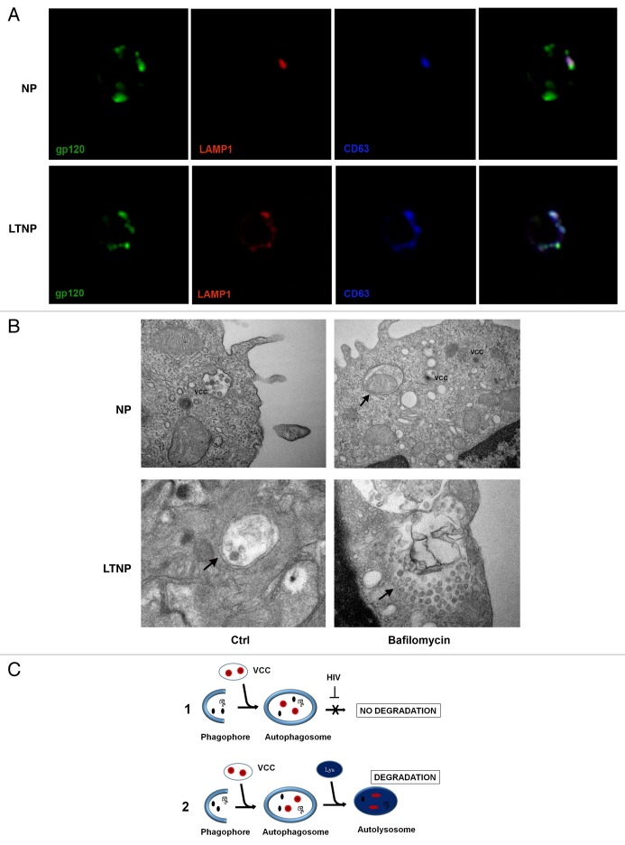
Figure 5. HIV-1 particles inside AV. (A) Confocal microscopy immunolocalization of the HIV-1 protein gp120 (green), LAMP1 (red) and the tetraspanin CD63 (blue) on PBMC from NP and LTNP. (B) Ultrastructural images of PBMC from NP and LTNP patients showing VCCs that carried viral particles or viral components. An AV containing a mitochondrion was visible in bafilomycin A1-treated PBMC from NP (arrow). Control cells from LTNP showed a double-membrane AV containing viral particles or viral components (arrow). Bafilomycin-A1-treated LTNP PBMC showed a very large AV containing undigested material and viral particles or viral components (arrow). (C) Hypothetical mechanism of HIV-1 removal by autophagy. HIV-1 particles (red), budding into VCC, or viral components (black), can be captured by autophagosomes. Then the viral components degradation can be blocked by viral-specific protein/s in NP (C1). In contrast, HIV-1 components can get digested in autolysosomes in HIV-1 controllers (C2). Original magnification: (A) 63×; (B) NP Ctrl 30,000×, NP bafilomycin A1 30,000×, LTNP Ctrl 50,000×, LTNP bafilomycin A1 50,000×.

An official website of the United States government
Here's how you know
Official websites use .gov
A
.gov website belongs to an official
government organization in the United States.
Secure .gov websites use HTTPS
A lock (
) or https:// means you've safely
connected to the .gov website. Share sensitive
information only on official, secure websites.
