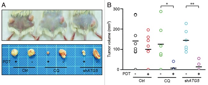
Figure 6. Inhibition of autophagy enhanced the antitumorigenicity of PDT in a xenograft model. (A) A total of 1 × 10³ viable PROM1/CD133+ PCCs, pretreated with CQ (10 μM), ATG5 shRNA lentivirus, or PDT (1.3 J/cm2), either alone or in the indicated combinations, were implanted subcutaneously into both flanks of NOD/SCID mice. Representative pictures of each treatment group. Mice were sacrificed 12 wk after implantation. (B) Tumor sizes were measured at the end of the experiment. Data are presented as dot plots, and the short black lines indicate mean tumor size. Tumor volume was calculated as (L * W2)/2, where L is the length and W is the width of the tumor. The tumors in CQ-treated and ATG5-silenced groups were significantly distributed in lower tumor volume ranges with PDT compared with without PDT. *P < 0.05; **P < 0.01.

An official website of the United States government
Here's how you know
Official websites use .gov
A
.gov website belongs to an official
government organization in the United States.
Secure .gov websites use HTTPS
A lock (
) or https:// means you've safely
connected to the .gov website. Share sensitive
information only on official, secure websites.
