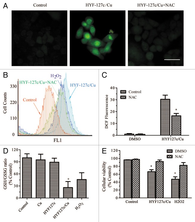
Figure 3. HYF127c/Cu induces cell death through oxidative stress. (A) Cells were treated with DMSO, 5 μM HYF127c/Cu or an additional 5 mM NAC treatment for 12 h. After incubation with 10 μM H2DCFDA, cells were washed and examined by fluorescence microscopy. Scale bar: 20 μm. (B) Cells were treated with the indicated compounds for 12 h. After incubation with 10 μM H2DCFDA, cells were washed and examined by flow cytometry. (C) Average fluorescence intensity from DCF. (D) Cells were treated with DMSO, 5 μM Cu, 5 μM HYF127c, 5 μM HYF127c/Cu, or 5 μM H2O2 for 12 h. GSH and GSSG were measured with a microplate reader. The relative ratio is shown as indicated (n = 3, *P < 0.05). (E) The relative ratio of cellular viability in cells treated with combinational compounds as indicated (n = 3, *P < 0.05).

An official website of the United States government
Here's how you know
Official websites use .gov
A
.gov website belongs to an official
government organization in the United States.
Secure .gov websites use HTTPS
A lock (
) or https:// means you've safely
connected to the .gov website. Share sensitive
information only on official, secure websites.
