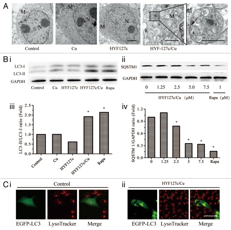
Figure 7. HYF127c/Cu induces autophagy in HeLa cells. (A) Electron microscopy images showing extensive cytoplasm vacuolization enclosed in a double membrane in HYF127c/Cu-treated HeLa cells. Electron microscopy image of an untreated cell is also shown for comparison. The double membrane of the autophagic vacuoles is indicated by a black arrow. N, nucleus; M, mitochondrion. Scale bar: 0.5 μm. (B) Conversion of LC3-I to LC3-II (i and iii) or degradation of SQSTM1 (ii and iv) in HYF127c/Cu-treated cells. HeLa cells were incubated with DMSO, 5 μM Cu, 5 μM HYF127c, 5 μM HYF127c/Cu or 1 μM rapamycin (control) and the amount of endogenous LC3-II proteins or SQSTM1 was analyzed by immunoblot. (C) The fluorescence images showing colocalization of lysosomes and autophagosomes in HYF127c/Cu-treated cells (ii) compared with the control (i). EGFP-LC3-transfected HeLa cells were stained with LysoTracker Red after 20 μM HYF127c/Cu treatment for 12 h. Scale bar: 20 μm.

An official website of the United States government
Here's how you know
Official websites use .gov
A
.gov website belongs to an official
government organization in the United States.
Secure .gov websites use HTTPS
A lock (
) or https:// means you've safely
connected to the .gov website. Share sensitive
information only on official, secure websites.
