Figure 6.
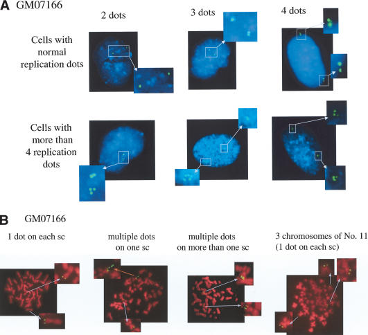
NBS deficiency leads to rereplication of DNA sequences near replication initiation sites. (A) Representative FISH analysis performed on interphase NBS-deficient cells (GM07166). Fixed cells were hybridized with a BAC probe corresponding to a segment covering the β-globin locus (clone RP11-645I8), and the hybridization images appear as green spots. This cloned genomic segment contains a putative replication origin (Kitsberg et al. 1993; Aladjem et al. 1995). Chromosomal DNA was stained with DAPI (blue). Examples of GM07166 cells that contain two, three, four, or more dots at the β-globin locus are shown. (B) FISH of metaphase nuclei of GM07166 cells. Cells were treated with colchicine to select for metaphase cells, and metaphase spreads were prepared. The fixed cells were hybridized with the above-noted β-globin probe (green), and chromosomes were stained with PI (red). FISH-positive segments of chromatids are identified with arrows in magnified copies of the relevant segments of each spread. In the various images are depicted GM07166 nuclei containing two pairs of β-globin dots (normal; one on each sister chromatid [sc]); multiple dots on one sc; multiple dots on more than one sc; and three pairs of dots each on a chromosome (indicating the presence of three No. 11 chromosomes). In some cases, the highly packed metaphase chromosomes likely “opened up” in certain locations during preparation, possibly allowing DNA to loop out, which, in turn, led to FISH signals extending out from the main body of the chromosome. An example is shown in the “multiple dots on one sc” and is marked with a red arrow. Here, the amplification of the β-globin DNA sequence of interest in the indicated chromatid is reflected by the presence of multiple FISH dots in a linear array extending out from the relevant chromatid.

