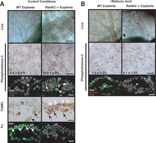
Figure 4.
In vitro Raldh2-/- yolk sac vascular phenotype and rescue with RA. (A) Wild-type (WT; left) and Raldh2-/- (right) explants after 48 h in control conditions in whole embryo culture recapitulated our in vivo findings as Raldh2-/- yolk sac failed to remodel (top row; bar, 100 μm). Dashed lines highlight vascular patterning in yolk sac. Whole-mount immunostaining of yolk sac tissues (second row; bar, 100 μm) with anti-phosphohistone-3 demonstrated a twofold increase in the mitotic index of endothelial cells in Raldh2-/- explants compared with wild type (WT), also corroborating in vivo findings. Furthermore, immunofluorescence (third row; bar, 25 μm) to colocalize phosphohistone-3 (green) with VE-Cad (red) demonstrated that the proliferating cells were predominantly endothelium (arrows). TUNEL assay of yolk sac sections (fourth row; bar, 25 μm) revealed a 10-fold increase in apoptosis in extraembryonic visceral endoderm (VE, white arrows) of Raldh2-/- explants. The level of apoptosis in mesoderm (M, black arrows) of Raldh2-/- explants was similar to that of wild type. Detection of Fn protein by immunofluorescence (bottom row; bar, 25 μm) demonstrated decreased Fn deposition specifically between the visceral endoderm and mesoderm layers (arrows) in Raldh2-/- explants. (B) Addition of 3 ng/mL RA to culture media restored vascular remodeling in yolk sacs of Raldh2-/- explants (top) and had no effect on wild-type development. Whole-mount (middle row; bar, 100 μm) immunostaining with antiphosphohistone-3 and immunofluorescence (bottom row; bar 25 μm) with antiphosphohistone-3 (green) and VE-cad (red) revealed that RA restored endothelial cell cycle control in Raldh2-/- explants to wild-type level.

