Figure 6.
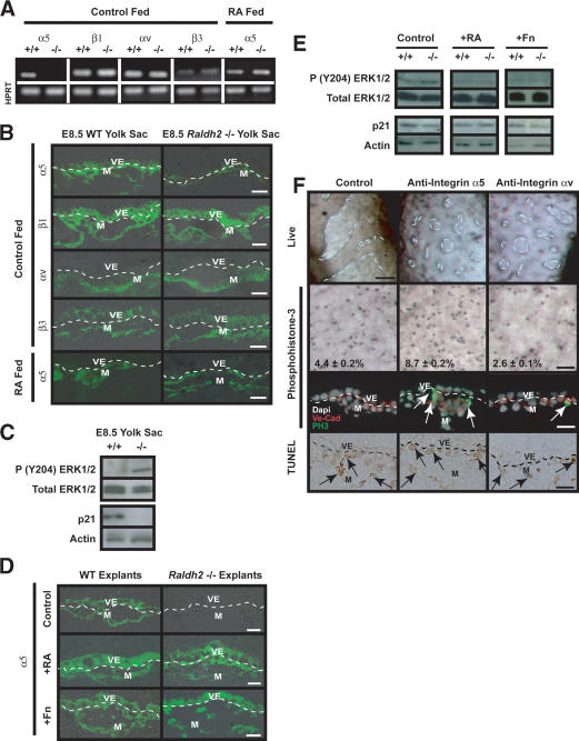
Endothelial cell proliferative control is regulated by extracellular matrix proteins via integrin α5 and integrin αv. (A) sqRT-PCR analysis of E8.5 Raldh2-/- and wild-type (WT) yolk sacs revealed down-regulation of integrin α5 transcript, but no change in β1, αv, or β3 levels. Feeding RA to pregnant females from E7.5-E8.5 restored α5 transcript levels in Raldh2-/- yolk sacs. HPRT was used as an internal control. (B) Immunofluorescence demonstrated that integrin α5 protein is decreased in control fed E8.5 Raldh2-/- yolk sacs compared with wild type (WT; top row), but protein expression was restored by feeding RA to pregnant females from E7.5-E8.5 (bottom row). There were no differences in protein levels of β1 (second row), αv (third row), or β3 (fourth row) in E8.5 Raldh2-/- yolk sacs compared with wild type. (C) Western blot analyses of E8.5 Raldh2-/- and wild-type (WT) yolk sac protein demonstrated increased phosphorylation of ERK1/2 at Tyr 204 (top row) in mutant yolk sacs compared with wild type. Conversely, protein levels of p21 (third row) were decreased in Raldh2-/- yolk sacs. Total ERK (second row), and actin (bottom row) were used as loading controls. (D) Immunofluorescence of embryo culture sections revealed that integrin α5 protein, which was decreased in control treated Raldh2-/- explants (top row), was restored by treatment with 3 ng/mL RA (middle row) and 100 μg/mL Fn (bottom row). (E) Western blot analyses of yolk sacs from embryo culture explants demonstrated that phosphorylation of ERK1/2 (Tyr 204), which were increased in untreated Raldh2-/- mutants, were restored to wild-type levels by treatment with 3 ng/mL RA and 100 μg/mL Fn. Protein levels of p21 were decreased in untreated Raldh2-/- explants but restored by treatment with RA. (F) Blocking antibodies to integrin α5 (middle column) or integrin αv (right column) inhibited vascular remodeling in 40% of wild-type explants in whole embryo culture (top row; bar, 100 μm). Whole-mount immunostaining (second row; bar, 100 μm) with antiphosphohistone-3 and immunofluorescence (third row; bar, 25 μm) with antiphosphohistone-3 (green) and VE-Cad (red) revealed that blocking integrin α5 signaling increased endothelial cell proliferation. Conversely, inhibiting integrin αv resulted in decreased proliferation. TUNEL assay (bottom row) demonstrated no differences in apoptosis in the mesoderm (arrows) and visceral endoderm in control and α5 or αv antibody treated explants.

