
Figure 1.
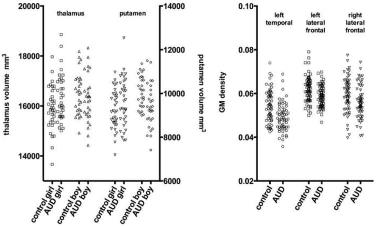
The subcortical volumes for the thalamus and putamen for each subject is shown on the left, grouped by alcohol status and sex, showing that AUD girls had larger volumes and AUD boys had smaller volumes. The average GM density within the left temporal/parietal/frontal, left lateral frontal, and right lateral frontal clusters is shown for each participant on the right, grouped by alcohol status, illustrating the generally smaller GM density in the AUD subject of each pair matched on age, sex, and acquisition protocol.