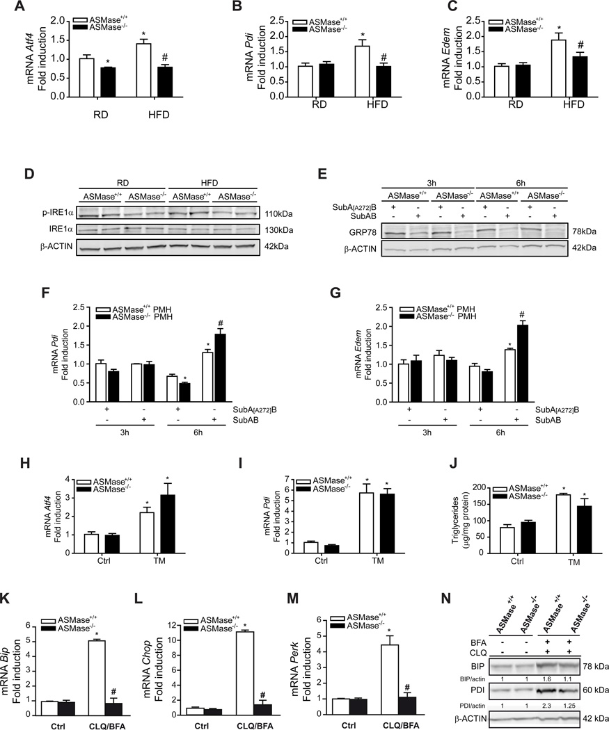
Figure 2. Resistance of ASMase−/− mice to HFD-mediated ER stress.
Expression of ER stress markers, (A) Atf4, (B) Pdi and (C) Edem, and (D) p-IRE1αινλιωερ from mice fed RD or HFD. Moreover, PMH were treated with SubAB to examine GRP78 degradation (E), and the expression of Pdi (F) and Edem (G). ASMase+/+ mice or ASMase−/− mice were treated with tunicamycin (TM) examining the expression of Atf4 (H), Pdi (I) and liver TG levels (J). PMH were cultured in the presence of chloroquine (CLQ) and brefeldinA (BFA) and samples processed for (K) BiP, (L) Chop, (M) Perk expression and western blot (N). Results are expressed as mean ± SD (n=5–7 mice). A–C: *p< 0.05 vs. RD-fed ASMase+/+ mice; F–G: *p<0.05 vs. SubA[A272]B-treated ASMase+/+ pmh, #p<0.05 vs SubAB-treated ASMase+/+ PMH; H–J: *p<0.05 vs. control.

