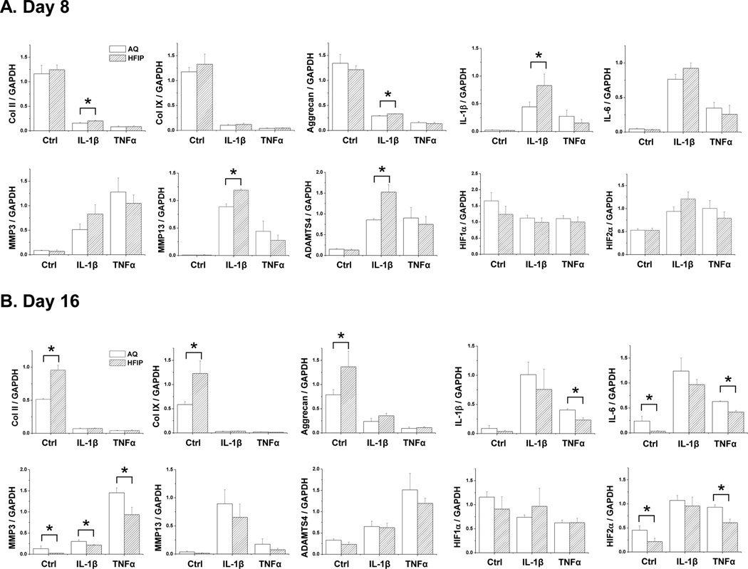
Fig. 2. qRT-PCR Gene expression analysis of chondrocytes grown in AQ and HFIP silk scaffolds.
Bovine articular chondrocytes were grown in AQ and HFIP silk scaffolds for 8 or 16 days with the following treatment: control (no cytokine treatment), IL-1β (10ng/ml) and TNFα (10ng/ml). The expression of the following genes was analyzed: collagen II (Col II), collagen IX (Col IX), aggrecan, endogenous IL-1β, IL-6, MMP3, MMP13, ADAMTS4, HIF1α and HIF2α. For each treatment, results from three independent samples are shown. (A) Results from Day 8 cultures. (B) Results from Day 16 cultures. All gene expression levels were normalized to GAPDH. Data present mean ± SD. *p<0.05.

