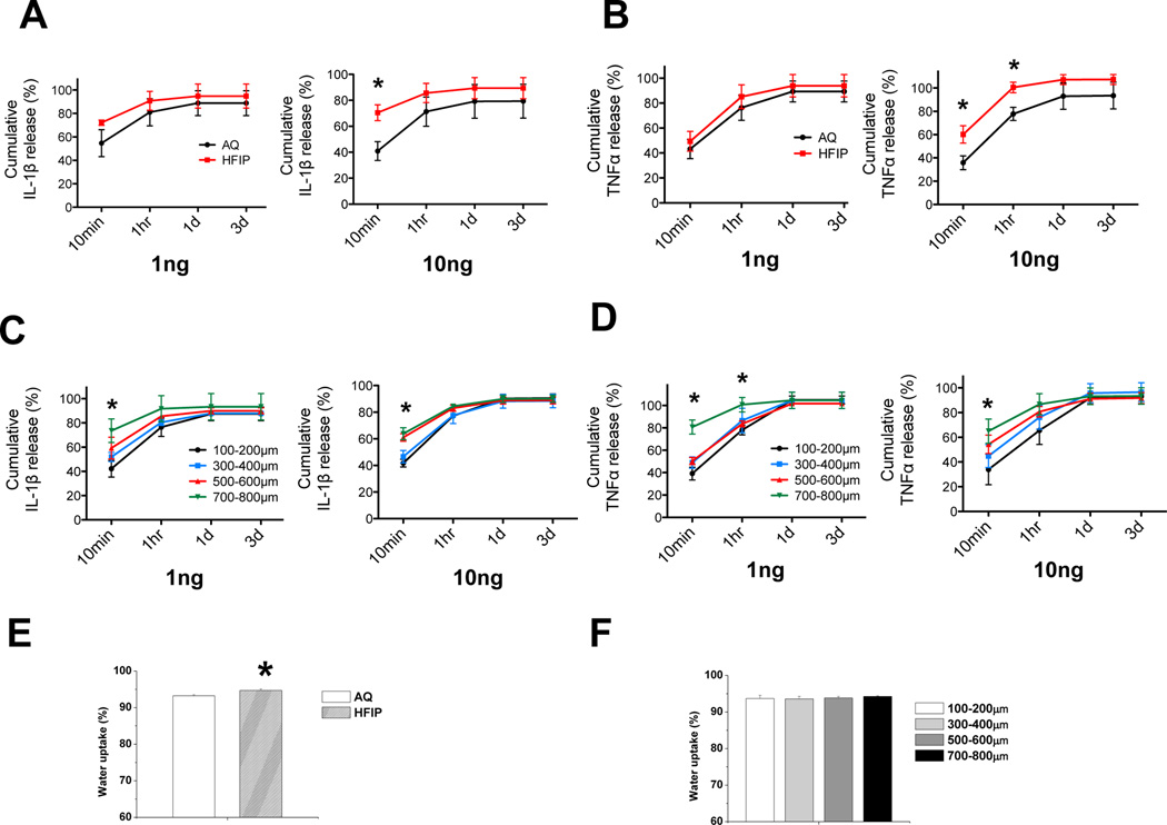
Fig. 5. Evaluation of kinetics of scaffolds in releasing pro-inflammatory cytokines and water uptake properties of the scaffolds.
(A–D) Two different amounts of pro-inflammatory cytokines IL-1β or TNFα (1 and 10ng) were loaded onto empty prewetted AQ and HFIP scaffolds, as well as HFIP with different pore sizes, and scaffolds were placed into the culture medium. At 4 different time points: 10min, 1hr, 1 day, and 3 days, the amount of IL-1β or TNFα leached from the scaffolds into the medium were evaluated by ELISA. Percent release was calculated as the ratio of the amount of cytokines in the medium to the total amount of cytokines loaded onto the scaffolds. Percent cumulative release was calculated as the ratio of cumulative amount of cytokines in the medium at each time point (i.e. sum of mass of cytokines at each time point and all prior time points) to the total amount of cytokines loaded onto the scaffolds. (A) Percent release and cumulative release analyses of IL-1β from AQ and HFIP silk scaffolds at each time point. (B) Percent release and cumulative release analyses of TNFα from AQ and HFIP silk scaffolds at each time point. (C) Percent release and cumulative release analyses of IL-1β from HFIP silk scaffolds with different pore sizes at each time point. (D) Percent release and cumulative release analyses of TNFα from HFIP silk scaffolds with different pore sizes at each time point. (E–F) Water uptake properties of AQ, HFIP, and HFIP silk scaffolds with different pore sizes were examined. (E) Water uptake (%) analyses of AQ and HFIP silk scaffolds. (F) Water uptake (%) analyses of HFIP silk scaffolds with different pore sizes. Data present mean ± SD. *p<0.05.

