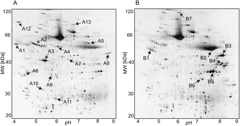
Figure 2. Details of the 2-D patterns of placental proteins incubated with HTRA1.
The placenta proteins were incubated for 4 h at 37°C without (A) or with the protease HTRA1 (B). Numbers indicate protein spots that were subjected to tryptic in-gel digestion and LC-MS (A = disappeared spots, B = appeared spots). Details and accession no of the substrates can be found in Table 1. The shown images are representative of four independent experiments, one in the presence of the inhibitor Timp-1 and one in the presence of an inhibitor cocktail (Fig. S2 and S3).

