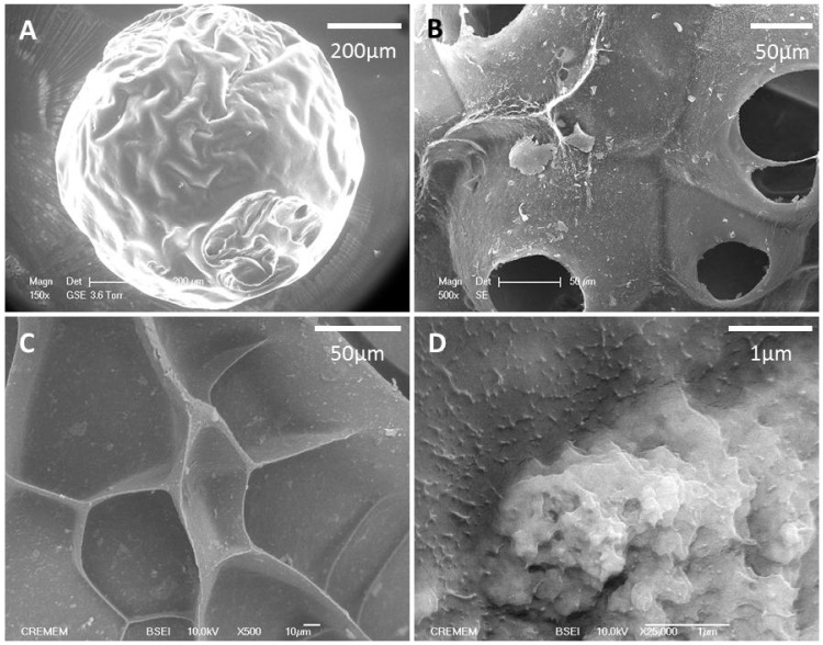
Figure 1. The morphology of microspheres was analyzed by scanning electron microscopy (A).
After rehydration in PBS, porosity of hydrated scaffolds was observed with an Environmental Scanning Electron Microscopy (B). Back-scattered Electron Microscopy images of dry beads show pores and overall surface properties (C) and the distribution of nHA aggregates within the structure (D).

