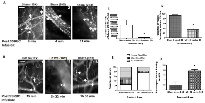
Figure 4. The MEK inhibitor U0126 abrogates SSRBC adhesion and vasoocclusion in vivo.
Nude mice implanted with dorsal skin-fold window chambers were injected with murine TNFα. Four hours later, intravital microscopic observations of post-capillary venules using 10× and 20× magnifications were conducted through the window chamber immediately after infusion of fluorescently-labeled sham- (A; panels 1, 2 and 3) or U0126-treated (B; panels 1, 2 and 3) human SSRBCs (n = 5). SSRBC adhesion and vasoocclusion are indicated with arrows. While sham-treated SSRBCs adhered markedly to venule walls promoting vasoocclusion, U0126 treatment of SSRBCs significantly reduced SSRBC adhesion and stasis. Scale bar = 50 µm. C. Video frames showing vessel segments were used to quantify adhesion in venules of animals occupied by SSRBCs (n = 5 for each treatment). Adhesion of fluorescently labeled sham-treated SSRBCs (sham-treated SS) and U0126-treated SSRBCs (U0126-treated SS) observed in all vessels recorded presented as fluorescence intensity of adherent SSRBCs (pixels). D–F. The values of at least 35 segments of vessels were analyzed and averaged among groups of animals (n = 5) to represent percentage of vessels occupied by adherent SSRBCs (D); percentage of vessels with normal blood flow, slow blood flow and no blood flow (E); and percentage of normal flowing vessels (F). Error bars show SEM of 5 different experiments for each treatment condition. *: p = 0.0103 (C) and p<0.0001 (D and F) compared to sham-treated SS regardless of the vessel diameter within the ranges specified.

