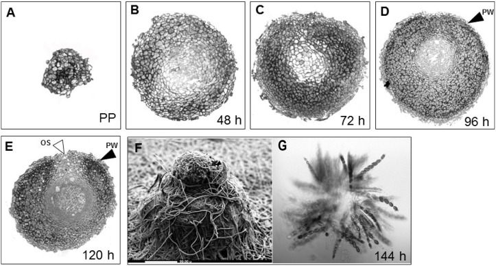
Figure 1. Sexual development of N. crassa.
Cross sections of developing perithecia, from A) the protoperithecium (PP) through a time course of (B) 48 h, (C) 72 h, (D) 96 h, and (E) 120 h after fertilization. These images illustrate the development of several cell layers within the fruiting body such as the perithecium wall (PW, black arrowheads), composed of thick-walled cells, as well as the initial stages of formation of asci and ascospores and the ostiole (OS, white arrowheads) through which spores are released in a later stage. After (F) 144 h, the perithecium including its beak is fully developed, as shown by scanning electron microscopy (SEM). (G) A squash mount of a mature fruiting body, showing asci and ascospores.

