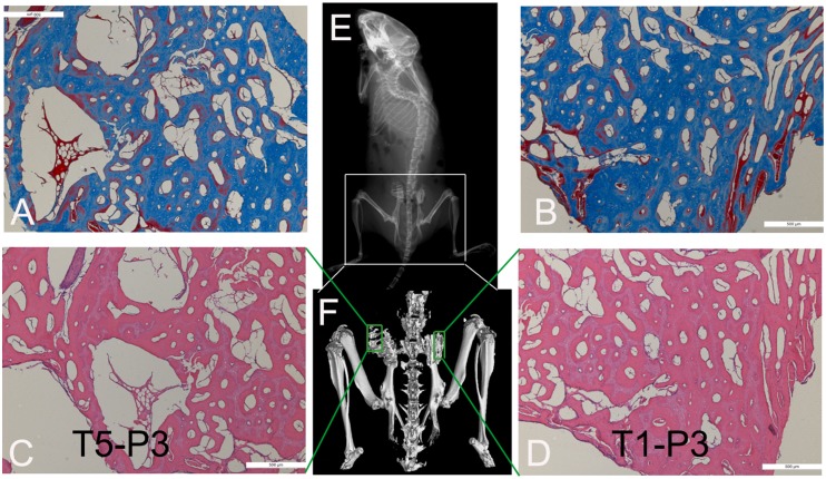
Figure 6. Osteogenic evaluation in vivo of TEB containing T5-P3 hUCMSCs (A and C) and T1-P3 hUCMSCs (B and D).
X-ray (E), micro-CT (F), and staining of Masson (A and B) and HE (C and D)were applied to demonstrat new bone formation and neoformative blood vessels in hibateral implanted TEB. Scale bars: 500 um in A–D.

