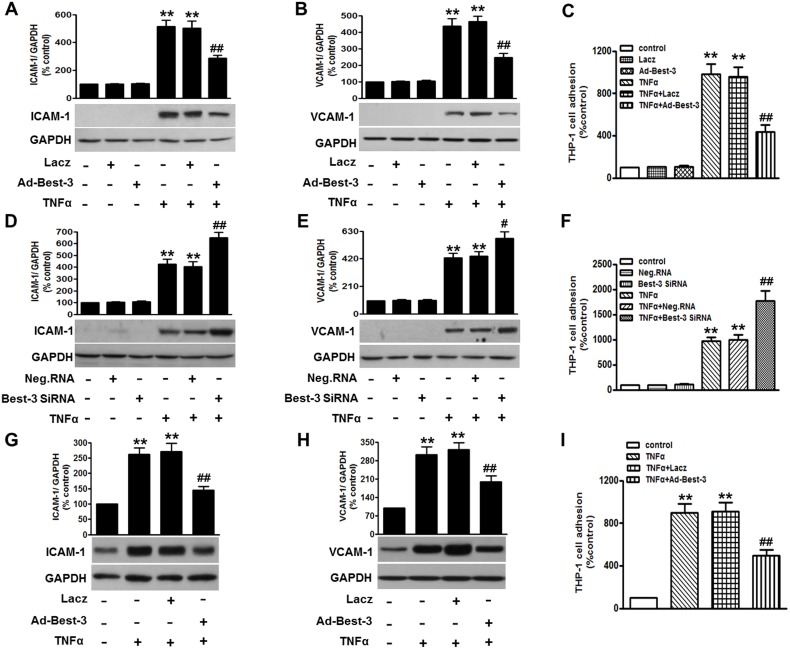
Figure 2. Best-3 ameliorated TNFα-induced inflammatory response in endothelial cells.
(A, B) HUVECs were transfected with Lacz or Ad-Best-3 for 48 h prior to TNFα treatment for 24 h. ICAM-1 (A) and VCAM-1 (B) were examined by western blot, respectively. (C) after treatment mentioned in (A, B), adhesion of VibrantDiO-labeled THP-1 to HUVECs were analyzed. (D, E) HUVECs were transfected with negative siRNA (Neg. RNA) or Best-3 siRNA for 48 h prior to TNFα incubation. ICAM-1 (D) and VCAM-1 (E) were detected by western blot, respectively. (F) after treatment mentioned in (D, E), adhesion of THP-1 to HUVECs was analyzed. (G, H) western blot detection of ICAM-1 (G) and VCAM-1 (H) expressions in MAECs isolated form mice after treatment mentioned in method section. (I) adhesion of THP-1 to MAECs was analyzed. All data are presented as mean ± SEM. **P<0.01 vs. control, #P<0.05, ##P<0.01 vs. TNFα alone, n = 6.

