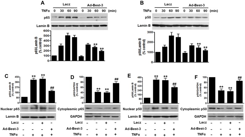
Figure 4. Best-3 repressed TNFα-induced NF-κB activation in endothelial cells.
(A, B) HUVECs were infected with Lacz or Ad-Best-3 for 48 h, and then incubated with TNFα (10 ng/ml) for different times as indicated. Nuclear fractions were isolated and detected by western blot using p65 (A) and p50 (B) antibodies. **P<0.01 vs. similarity treated control, n = 4. (C–F) nuclear and cytoplasmic fractions of MAECs isolated from mice after treatment mentioned in method section were analyzed by western blot to detect the expressions of p65 (C, D) and p50 (E, F). **P<0.01 vs. control, ##P<0.01 vs. TNFα alone, n = 6.

