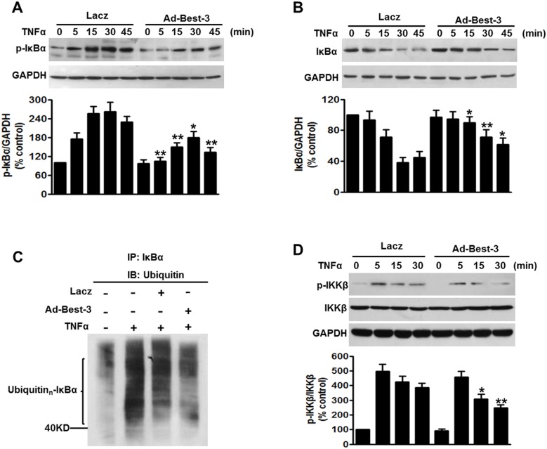
Figure 5. Best-3 suppressed IKKβ/IκBα pathway to inhibit inflammation.
(A, B) HUVECs were infected with Lacz or Ad-Best-3 for 48 h, and then incubated with TNFα (10 ng/ml) for different times as indicated. Cell lysates were subjected to western blot analysis using p-IκBα (A) and IκBα (B) antibodies. *P<0.05, **P<0.01 vs. similarity treated control, n = 4. (C) western blot analysis of ubiquitinated IκBα in HUVECs treated with Lazc or Ad-Best-3 in the presence of TNFα for 30 min. Cell lysates were immunoprecipitated with IκBα antibody and immunoprecipitated proteins were blotted with ubiquitin antibody to reveal ubiquitination of IκBα. (D) western blot analysis of p-IKKβ and IKKβ of HUVECs treated with Lacz or Ad-Best-3 for 48 h in the presence of TNFα for different times as indicated. *P<0.05, **P<0.01 vs. similarity treated control, n = 4.

