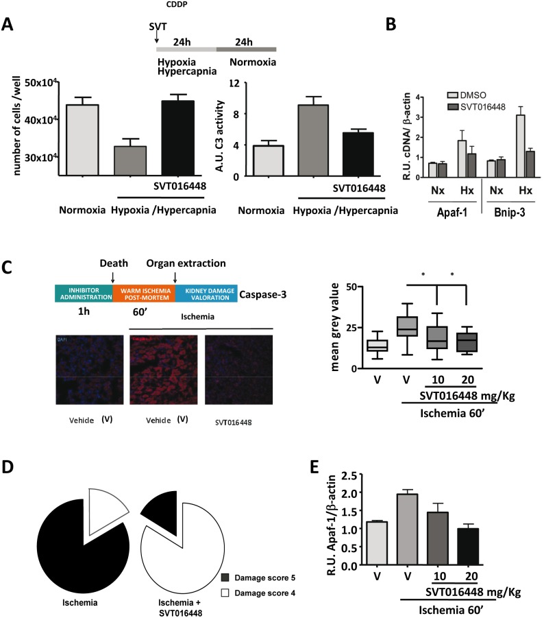
Figure 4. Apaf-1 inhibitors provide protection in an in vivo animal ischemic induced acute kidney injury model.
(A) Cell survival and caspase-3 activity in cell lysates from LLC-PK-1 cells measured after 24 h hypoxia/hypercapnia plus 24 h normoxia in the absence or presence of Apaf-1 inhibitor (10 µM) (mean ± SD; n = 3). (B) Transcriptional regulation of Apaf-1 and Bnip-3 were analyzed by RT-qPCR in LLC-PK-1 cells. mRNA levels were compared in hypoxia/hypercapnia (Hx) versus normoxia (Nx) conditions after SVT016448 treatment (mean ± SD; n = 3). (C) Intraperitoneal injection of SVT016448 (10 mg/Kg) decreases caspase activity in a renal hot ischemia in vivo model. A representative image of the immunofluorescence of active caspase-3 (left panel) and mean grey value quantification (right panel) (mean ± SD, n = 5). Asterisks represent significant differences relative to ischemia treatment as determined by one-way ANOVA test with Bonferroni’s multiple comparison post-test (*p<0.05). (D) Damaged tissue decreases in the presence of the Apaf-1 inhibitor, as determined by histopathological evaluation. (E) SVT016448 treatment-dependent decrease in Apaf-1 mRNA levels, as analyzed by RT-qPCR.

