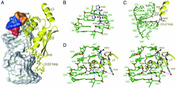
Fig. 3.
Interactions between Rnt1p dsRBD and snR47h RNA. Lowest energy structure is shown. (A) Solvent-accessible surface of the RNA showing the major and minor grooves with the AGAA tetraloop nucleotides colored red, blue, and orange, and the protein in yellow ribbon. Protein side chains that interact with the RNA are shown as sticks. (B–D) Details of specific interactions of the protein with the minor groove (B), major groove (C), and tetraloop minor groove (stereoview) (D) are shown. Nucleotides are green, with phosphates and O2′ of interacting riboses in red. Protein side chains are sticks, with backbone as yellow ribbon. Direct and water-mediated protein–RNA H-bonds are indicated as orange and blue dashed lines, respectively.

