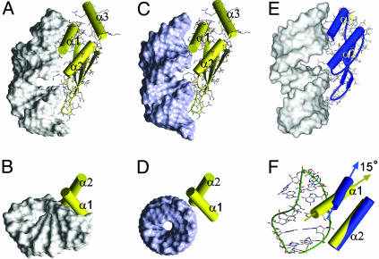
Fig. 5.
Comparison of positions of dsRBDs on RNA. Rnt1p dsRBD bound to snR47h (A and B) and modeled dsRNA (C and D). Side (A and C) and top (B and D) views are shown. (E) Side view of Xlrbpa dsRBD bound to dsRNA. (F) Comparison of Rnt1p (yellow) and Xlrpba (blue) α1 and α2 orientations on snR47h. The cylinder representations of the α-helices of Rnt1p (yellow) and Xlrpba (blue) dsRBDs were superimposed in F on their β-sheets, N termini of α2, and the β1–β2 loops. In A–E, the RNA is shown as solvent-accessible surface.

