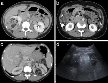
Figure 1.

Imaging appearances of angiomyolipomas. (a) Characteristic CT appearance of large right renal angiomyolipoma showing a heterogenous lesion with areas of fat density and a small lesion in the contralateral kidney. (b) Tiny exophytic cortical angiomyolipoma in the right kidney assigned a measurement of 5 mm (arrow). (c) & (d) CT and ultrasound appearances respectively of the same 40 mm angiomyolipoma.
