Fig. 4.
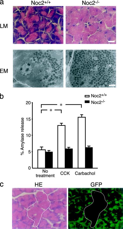
Histological and functional analysis of exocrine pancreas. (a) Histological analysis of exocrine pancreas. (Upper) Light microscopic (LM) analysis of acinar cells of exocrine pancreas stained with hematoxylin/eosin. (Lower) Electron microscopic (EM) analysis of acinar cells. Acinar cells in Noc2-/- mice are enlarged due to a remarkable accumulation of secretory granules throughout the cytoplasm. (Scale bars: 10 μmin Upper and 3 μmin Lower.) (b) Amylase secretion from exocrine pancreas in vitro. Amylase secretion was determined by the amount released into medium relative to total cellular content (expressed as percent amylase release) of pancreatic acinar cells. Open columns, Noc2+/+ mice; filled columns, Noc2-/- mice. Both CCK and carbachol stimulate amylase secretion in Noc2+/+ mice significantly, but neither stimulates amylase secretion in Noc2-/- mice [Noc2+/+ mice: 5.6 ± 0.9% (basal level), 13.0 ± 0.7% (CCK-stimulated amylase secretion), and 15.6 ± 0.8% (carbachol-stimulated amylase secretion), n = 12 for each; *, P < 0.0001, respectively; Noc2-/- mice: 4.9 ± 0.5% (basal level), 5.8 ± 0.5% (CCK-stimulated amylase secretion), and 6.2 ± 0.5% (carbachol-stimulated amylase secretion), n = 12]. Values are means ± SEM. (c) Histological analysis of exocrine pancreas of chimeric mice between GFP-Tg and Noc2-/- mice. Exocrine pancreas of the chimeric mice shows a mosaic pattern of mixed populations of GFP-positive acinar cells of normal appearance and GFP-negative acinar cells (circled by white line) having an overabundance of zymogen granules. GFP-positive and -negative cells originate from GFP-Tg and Noc2-/- mice, respectively. (Left)A section stained with hematoxylin/eosin. (Right) A section viewed under a fluorescent microscope. (Scale bar: 10 μm.)

