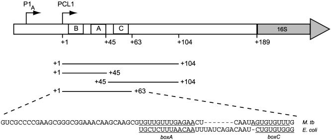
Fig. 1.
The M. tuberculosis rrnA leader and spacer region. The transcription start points of the two promoters, P1A and PCL1, are indicated with arrows, and the position of boxB (+2 to +19), boxA (+33 to +45), and boxC (+52 to +59) are indicated. The probes used in this study correspond to PCL1 transcripts and are shown with their 5′ and 3′ boundaries. The sequence of the M. tuberculosis (M. tb) rrn nut-like site (+1 to +63) are shown, and the boxA and boxC elements are underlined and aligned to the corresponding E. coli sequences.

