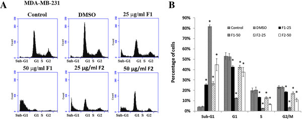
Figure 2.

Effect of F1 and F2 fractions on the cell cycle distribution in MDA-MB-231. (A) MDA-MB-231 cells were treated with 25 and 50 μg/ml of F1 or F2 fractions and control cells were treated with 0.5% DMSO for 48 hours, after which cells were stained with PI and analyzed for DNA content by flow cytometry. The sub-G1 peak is considered as the apoptotic portion. The results shown are representative of 3 independent experiments. (B) Bar graph shows the cell distributions of each phase of the cell cycle. Data are means ± SEM of three independent experiments. *denotes P < 0.05 versus DMSO group as measured by one-way ANOVA.
