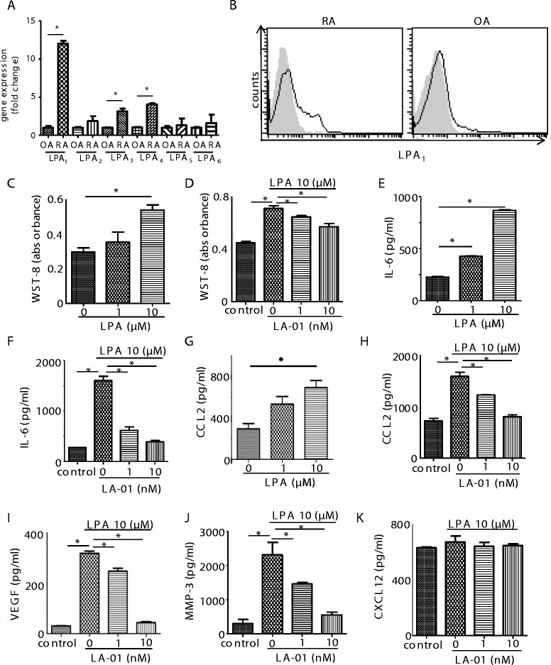
Figure 1.

Expression of lysophosphatidic acid receptors and the effect of lysophosphatidic acid receptor 1 on proliferation and production of inflammatory mediators in rheumatoid arthritis fibroblast-like synoviocytes. The expression levels of lysophosphatidic acid receptor 1 through 6 (LPA1–6) mRNA in fibroblast-like synoviocytes (FLSs) derived from the rheumatoid arthritis (RA) synovium (n = 10) were compared to those in FLSs from osteoarthritis (OA) synovium (n = 5) by real-time RT-PCR (A). Data were derived from samples from multiple individuals. Data are presented as the mean ± SEM. *P < 0.05 for RA vs OA. Cell surface expression of LPA1 on RA (n = 5) and OA (n = 3) FLSs was analyzed by flow cytometry (B). Filled histogram (gray): isotype control; open histogram (black line): LPA1. Representative histograms are shown. RA FLSs were cultured with lysophosphatidic acid (LPA) for 72 hours (C). FLSs were preincubated with an LPA1 inhibitor, LA-01, for 30 minutes,then stimulated with 10 μM LPA for 72 hours (D). Control: no stimulation with LPA. Cell proliferation was measured by using a cell counting kit (C) and (D). RA FLSs were cultured with LPA for 24 hours. Concentrations of interleukin 6 (IL-6) and chemokine (C-C motif) ligand 2 (CCL2) in the culture supernatant were measured by enzyme-linked immunosorbent assay (ELISA) (E) and (G). FLSs were preincubated with LA-01 for 30 minutes, then stimulated with 10 μM LPA for 24 hours. Concentrations of IL-6, CCL2, vascular endothelial growth factor (VEGF), matrixmetalloproteinase (MMP-3) and CXCL12 in the culture supernatant were measured by ELISA (F), and (H) through (K). Control: no stimulation with LPA. Data are presented as the means (±SEM) of one of three independent experiments analyzed in triplicate. *P < 0.05 vs control or LA-01 0 nM (C) through (K).
