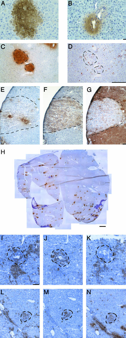
Fig. 1.
BMEL cells integrate and proliferate in the liver of Alb-uPA/SCID mice without having undergone cell fusion and elicit an immune reaction. (A and B) Immunohistochemistry revealing BMEL-GFP cells (brown) in the hepatic parenchyma (A) and as a bile duct-like structure (B). (C and D) Clusters of BMEL-GFP cells (C) are composed of proliferating cells as shown by the nuclear localization of the Ki67 antigen (D). Broken lines in this and the following figures delimit the fields of interest. GFP (E), H-2Kk (F), and H-2Dd (G) immunohistochemical analyses of adjacent serial sections are shown. Transduced BMEL cells do not all express GFP (E). H-2Kk reveals all BMEL cells injected (F) and H-2Dd identifies the host cells (G). The H-2Kk-positive field (F) is H-2Dd-negative (G), demonstrating that cell fusion, which would result in double-positive cells, is not involved. (H) A complete liver section after GFP staining reconstituted from 31 photographic images and illustrating the material used for quantitation detailed in Table 1. BMEL cell clusters are visible as brown spots, whereas the purple-stained regions are necrotic. (I–N) Infiltrations of immune cells are seen around BMEL cells after transplantation. BMEL H-2Kk-positive bile ducts (J and M) are surrounded by CD45+ cells (I and L), of which many are macrophages (antibody F/480) (K) and neutrophils (antibody Nimp-R14) (N). (Scale bars: A and B,10 μm; C–G, 100 μm; H, 1 mm; I–N,40 μm.)

