
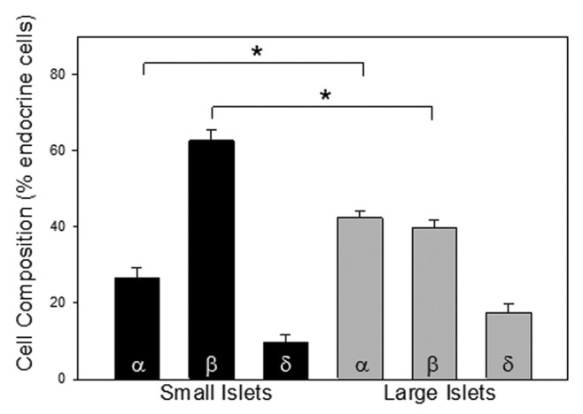
Figure 5. Cell composition. Islets were stained for α-cells (anti-glucagon), β-cells (anti-insulin) and δ-cells (anti-somatostatin). The percentage of each is plotted for small and large islets. There was a significantly greater percentage of α-cells and a lower percentage of β-cells in the large islets (n = 402 cells 6 donors, *p < 0.001).