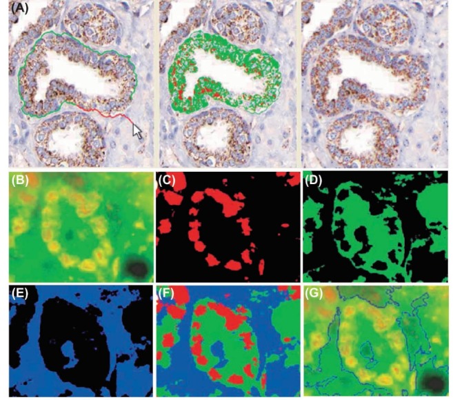
Fig. 7 .

Multiplexed and quantitative IHC using bioconjugated QDs. A) Prostate tissue specimens stained with traditional IHC and bioconjugated QDs. K-means clustering to segment QD-stained tissue image is highlighted by light green and light red colors. Panels B to G represent multiplexed QD-based IHC of the formalin fixed, paraffin embedded (FFPE) prostate tissue samples, and quantitative analysis of cancer biomarkers p53 and EGR-1. The blue color shows the tissue background. B) Original multicolor image. C) p53 protein stained red with QD655. D) EGR-1 protein stained green with QD565. E) Tissue background. F) Superimposed map of dominant markers and background. G) Automated boundary segmentation using level-set algorithms. IHC: immunohistochemistry. Data were adapted with permission from a study published by Xing et al.106
