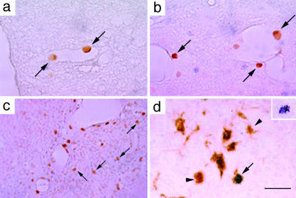
Fig. 1.
Localization and identification of HRP-labeled cells in Hexb-/- mice. In 3-month-old Hexb-/- mice, HRP-labeled cells were detected attached to the vascular wall in vessels in the spinal cord (a) and perivascular regions in the spinal cord (b). (c) In a 4-month-old Hexb-/- mouse, HRP-labeled cells were localized in the parenchyma of the thalamic nucleus. (d) An HRP-labeled cell in the thalamic nucleus of a 4-month-old Hexb-/- mouse was identified as Mac-1-positive (arrow). Arrowheads show Mac-1-positive cells. (Inset) A nickel-enhanced HRP-labeled cell. (Bars: 100 μmin a and b, 220 μmin c, and 60 μm in d.)

