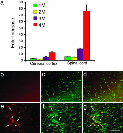
Fig. 2.
Expression of MIP-1α in Hexb+/+ and Hexb-/- mice. (a) The time course of relative MIP-1α mRNA expression levels in the cerebral cortex and spinal cord of Hexb-/- mice compared with age-matched Hexb+/+ mice. (b–g) Immunofluorescent staining of the spinal cord with MIP-1α and GFAP antibodies. (b–d) Control (Hexb+/+) sections. (e–g) Hexb-/- sections. b and e show MIP-1α immunostaining. c and f show GFAP immunostaining. d and g show double immunostaining for MIP-1α and GFAP. Note that the MIP-1α and GFAP double-positive astrocytes (arrows in e–g) are closely associated with vessels (*). (Bar: 60 μm.)

