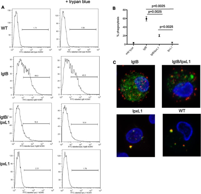
Figure 2.
Internalization of LOS modified NOMV by dendritic cells. Human monocyte-derived DC were co-cultured with 10 μg ml−1 FITC labelled NOMV for 4 h. DC were separated into 3 aliquots to distinguish internalized and surface adhered NOMV. One aliquot was left untreated and the second was treated with 0.4% trypan blue to quench FITC signal emitted from extracellular NOMVs. DC were then analysed by flow cytometry. The third aliquot was stained with To-Pro3 (blue) to stain the nucleus and anti-meningococcal serosubtype P1.7 antibody followed by Alex Fluor 568 goat anti-mouse to stain the surface adhered NOMV. Cells were then analysed by Confocal Microscopy. Data are shown from one representative donor (A and C) together with the summary of data from eight donors (B). Data are expressed as the mean and standard error of the mean and significance was determined by a paired t-test.

