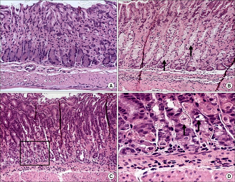
Figure 6.
Normal gastric mucosa (A) and mucous metaplasia and lymphoid aggregation in the gastric mucosa of Helicobacter pylori (H. pylori) infected C57BL/6 mice on a basal diet (B) and a high-salt diet (C, D) 4 weeks after inoculation (H&E stain); ×100 (A–C), ×200 (D). The glandular epithelium has been replaced by a hyperplastic, hypertrophic mucous epithelium (arrow and black square).

