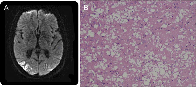
Figure. Brain MRI and brain pathology in Creutzfeldt-Jakob disease.

Axial diffusion-weighted image sequence (A) shows abnormal gyriform restricted diffusion in the bilateral parieto-occipital lobes. Cortical brain biopsy (B) shows gray matter with diffuse spongiform changes, neuronal dropout, and gliosis.
