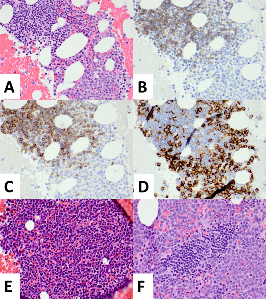
Figure 1.

A. Bone marrow clot section shows immature cells with monoblastic morphology (right lower corner) intermixed with mature-appearing lymphocyte (left upper corner), H & E, X40. B and C. The mature-appearing lymphocytes co-express CD5 and CD19, consistent with chronic lymphocytic leukemia (CLL), B-CD19 stain, C-CD5 stain, X40. D. CD11c stain is positive in monoblastic population and negative in CLL population, CD11c stain, x40. E. There is a residual monoblastic population, but CLL population is not observed, H & E, x40. F. CLL population is relapsed (center), demarcated from monoblastic population, H & E, X40.
