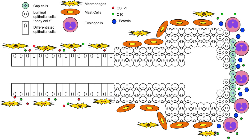
Figure 1. Model of immune cell localization at the terminal end buds during post-natal mammary ductal morphogenesis.
Immune cells including mast cells, eosinophils, and macrophages contribute to numerous effector functions during mammary ductal morphogenesis. Mast cells localize to the stromal regions surrounding proliferating terminal end buds (TEBs) (25). Next, macrophages are recruited to TEBs. After being recruited, macrophages migrate and localize to the neck of TEBs in response to CSF-1. Finally, eosinophils are recruited to the head of TEBs in response to expression of epithelial cell-secreted eotaxin (24). At the head of TEBs, eosinophils secrete the chemokine C10 which acts to recruit additional macrophages (36).

