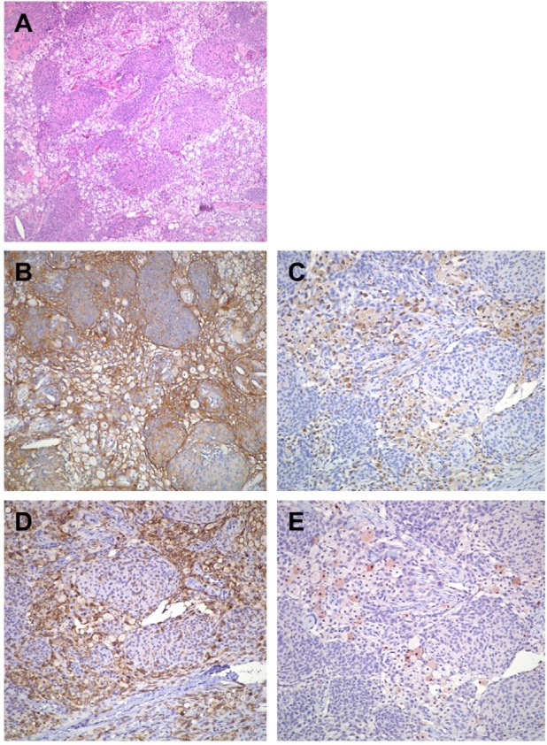
Figure 2.

The tumor exhibits atypical meningothelial proliferation with extensive and multifocal histiocytic infiltration. The histiocytic component constitutes approximately half of the entire tumor (A). Both meningothelial cells and histiocytic cells show immunoreactivity to EMA (B). The histiocytic cells are positive for CD68 (C) and CD4 (D). Scattered S100-positive histiocytes are seen throughout the tumor (E). (A: hematoxylin and eosin stain, 100×; B: EMA stain, 200×; C: CD68 stain, 200×; D: CD4 stain, 200×; E: S100 stain, 200×).
