
Figure 3.
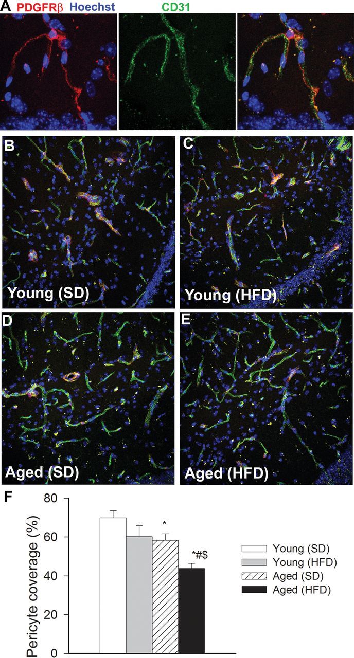
Obesity-induced changes in pericyte coverage of hippocampal capillaries in aging. (A) Representative confocal image showing perivascular localization of a platelet-derived growth factor receptor β (PDGFRβ) expressing pericyte (red) surrounding CD31 positive capillary endothelial cells (green) in the CA1 region of the mouse hippocampus. Hoechst 33342 was used for nuclear counterstaining. (B–E) Representative confocal microscopy analysis of PDGFRβ expressing pericyte coverage (red) of CD31 positive capillaries (green) in the CA1 region of the hippocampi of young SD-fed (B), young HFD-fed (C), aged SD-fed (D) and aged HFD-fed (E) animals. (F) Summary data showing obesity- and aging-dependent loss of pericyte coverage in the hippocampus (see Methods section for details). *p < .05 vs young mice (SD), # p < .05 vs aged mice (SD), $ p < .05 vs young mice (HFD).