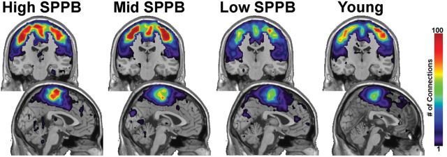
Figure 3.

First-order connections from somatomotor cortex. The majority of first-order connections are contained within the somatomotor regions. The images in the upper part of the figure are coronal slices through the somatomotor cortex. The lower images are midsagittal slices. Note that the color bar indicates the average number of connections across all participants in a group.
