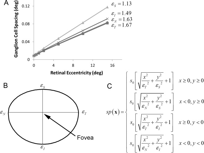
Figure 2.
Midget ganglion cell spacing (in degrees) in the human retina. (A) Ganglion cell spacing (1/square-root of density) in the four cardinal directions of the visual field, assuming one midget ganglion cell for each cone in the center of the fovea (which sets the y-intercept). This one “midget ganglion cell,” which can respond positively and negatively, represents an on-and-off pair of ganglion cells (data from Drasdo et al., 2007.) (B) To generate a ganglion cell mosaic, we assume that, in each quadrant, the contours of constant spacing fall on an ellipse. This specific contour shows the retinal locations at which the spacing between midget ganglion cells is twice what it is in the center of the fovea. Thus, in the upper vertical direction, the spacing doubles at about 1.1° of eccentricity, but in the horizontal directions, it does not double until about 1.6°. (C) The equation that defines the spacing function: s0 is the spacing in the center of the fovea, and εN, εT, εI, and εS are the eccentricities in the four cardinal directions where spacing reaches twice s0.

