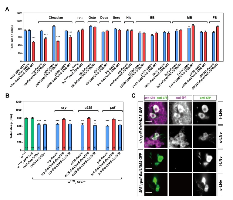
Figure 2. The sleep function of SPR is mapped to pdf neurons.
(A–B) Total sleep duration per day of virgin females of the indicated genotypes. Data are shown as means ± SEM. In (A), n = 16–64 for each bar. ***, p<0.001 for the comparison both Gal4 and UAS controls by Student's t test. In (B), number in bars indicates n of the tested flies. **, p<0.01, ***, p<0.001 for the comparison to w1118 controls by Student's t test. (C) pdf neurons express SPR. Confocal sections of the female brain of indicated genotypes stained by anti-SPR (magenta) and anti-GFP (green). Magenta and green channels are shown separately. Each brain hemisphere has four to five l-LNvs and four s-LNvs, most of which are labeled by anti-SPR (n = 6). Note that SPR expression is broad and not restricted to pdf neurons. Scale bar, 10 µm.

